Hello, I'm
Gia Jadick
PhD Candidate @ UChicago


Hello, I'm
PhD Candidate @ UChicago
Hello! I'm Gia Jadick. I am a medical physics PhD candidate at the University of Chicago, advised by Patrick La Rivière, where I develop computational methods for advanced X-ray and CT imaging. I successfully defended my PhD thesis on June 18, 2026, and am officially finalizing my degree this summer quarter.
My dissertation research unites traditional spectralimaging with propagation-basedphasecontrast through the lens of material decomposition. I study how and when conventional approximations in phase-contrast imaging break down, and I develop computational strategies for incorporating more general image acquisition models to improve basis material image reconstruction.
I am broadly interested in spectral photon-counting CT, phase contrast, inverse problems, and computational imaging. I have a strong technical foundation, building large-scale Python simulation and reconstruction tools that are efficiently GPU-parallelized with automatic differentiation compatibility. My recent work spans applications to high-resolution phase-contrast micro-CT of zebrafish, spectral photon-counting mammography for microcalcification morphology classification, multi-material decomposition for cardiac CT, and MV-kV dual-energy CT for radiotherapy imaging.
I graduated cum laude from Duke University in 2020 with a BS in physics and BA in political science. There, I researched scanner-specific CT simulation, mechanosensing of E. coli bacteria, and vintage saxophone mouthpiece acoustics.
My research code and related projects are publicly available on GitHub, with selected repositories linked below. My PhD studies are funded by the NSF Graduate Research Fellowship Program with additional support from the AAPM/RSNA Graduate Fellowship.
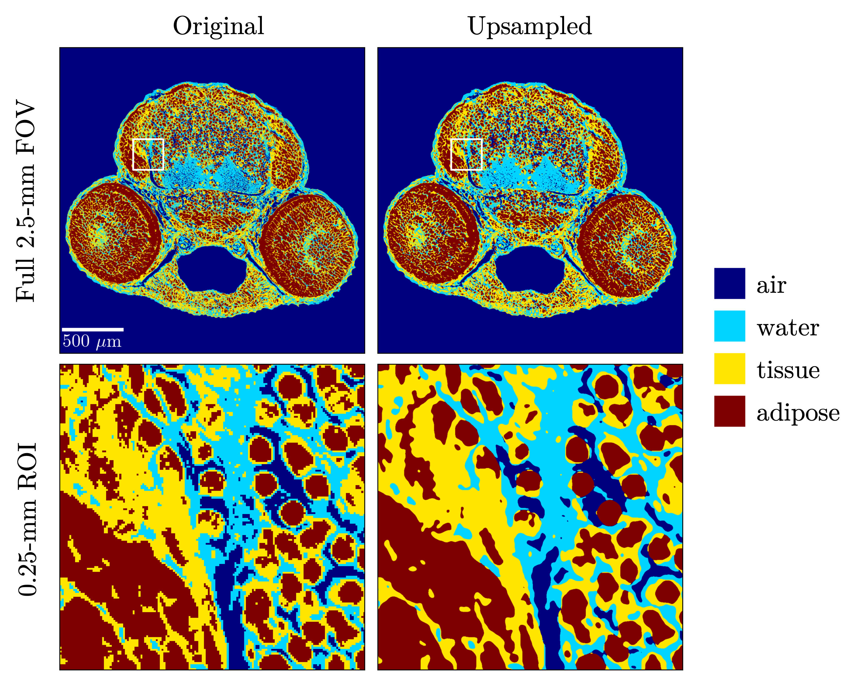
I developed a GPU-accelerated simulation framework for propagation-based phase-contrast synchrotron micro-CT, comparing the projection approximation with an auto-differentiable multi-slice forward model for more accurately including refractive effects within samples. Subsequent reconstructions were compared with our iterative method versus filtered-back projection and conventional phase retrieval. Manuscript in progress.
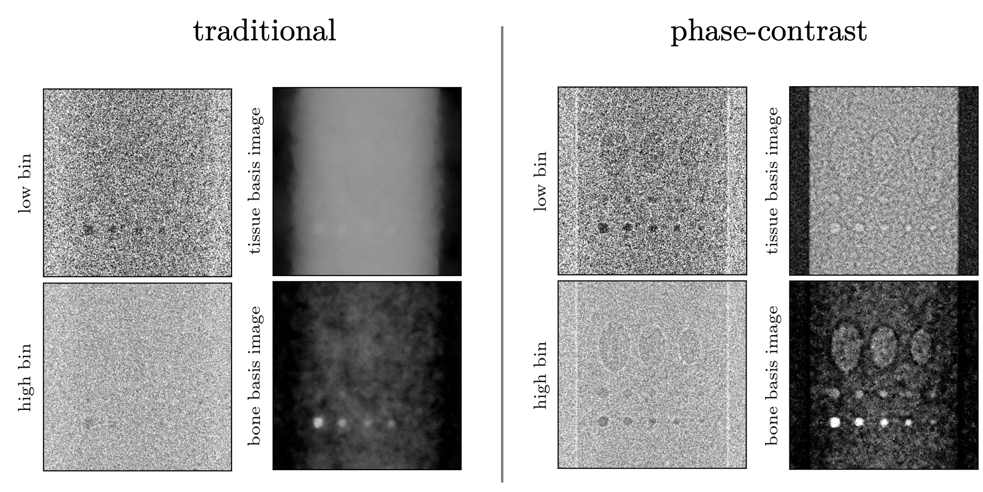
I expanded the multi-slice forward model to include a full polychromatic source spectrum and non-ideal energy-dependent system effects, developing a full spectral material decomposition method for X-ray phase-contrast imaging with energy-discrimating photon-counting detection. This was applied to a sensitivity analysis of model-based iterative material-decomposition phase-retrieval methods, quantifying the degree of improvement as more forward model elements were incorporated. Manuscript in progress.
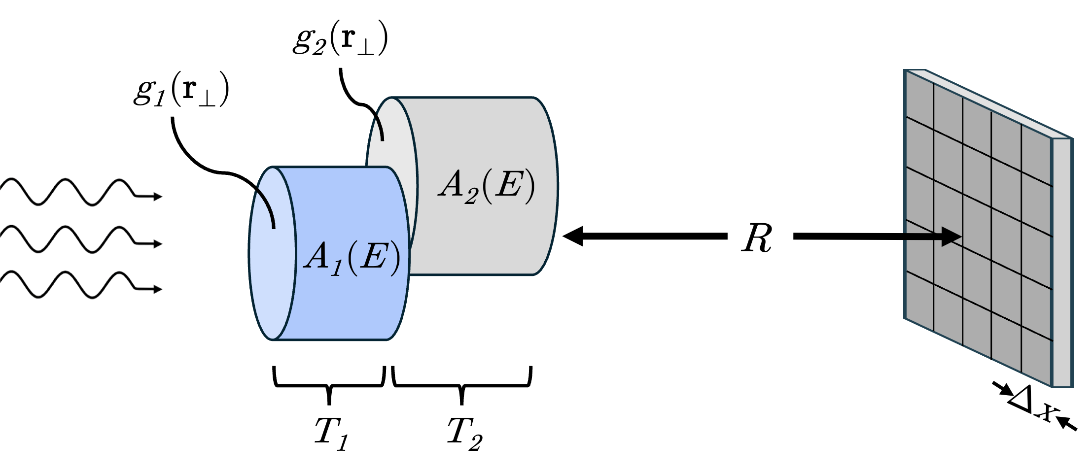
I developed a basis material Cramér–Rao lower bound (CRLB) framework for spectral propagation-based X-ray phase-contrast imaging, building various toy models to include the information of typical edge-enhancement effects. These models were applied to energy threshold optimization of polychromatic photon-counting detector measurements, including simulation validation with maximum likelihood estimation. Manuscript in progress.
I explored and quantified the potential utility of a novel dual-energy CT approach: combining a megavoltage (MV) and kilovoltage (kV) source in "MV-kV" imaging for use on radiotherapy systems that are conveniently already equipped with the two X-ray sources. Our publication was featured on the cover of the Journal of Medical Imaging.
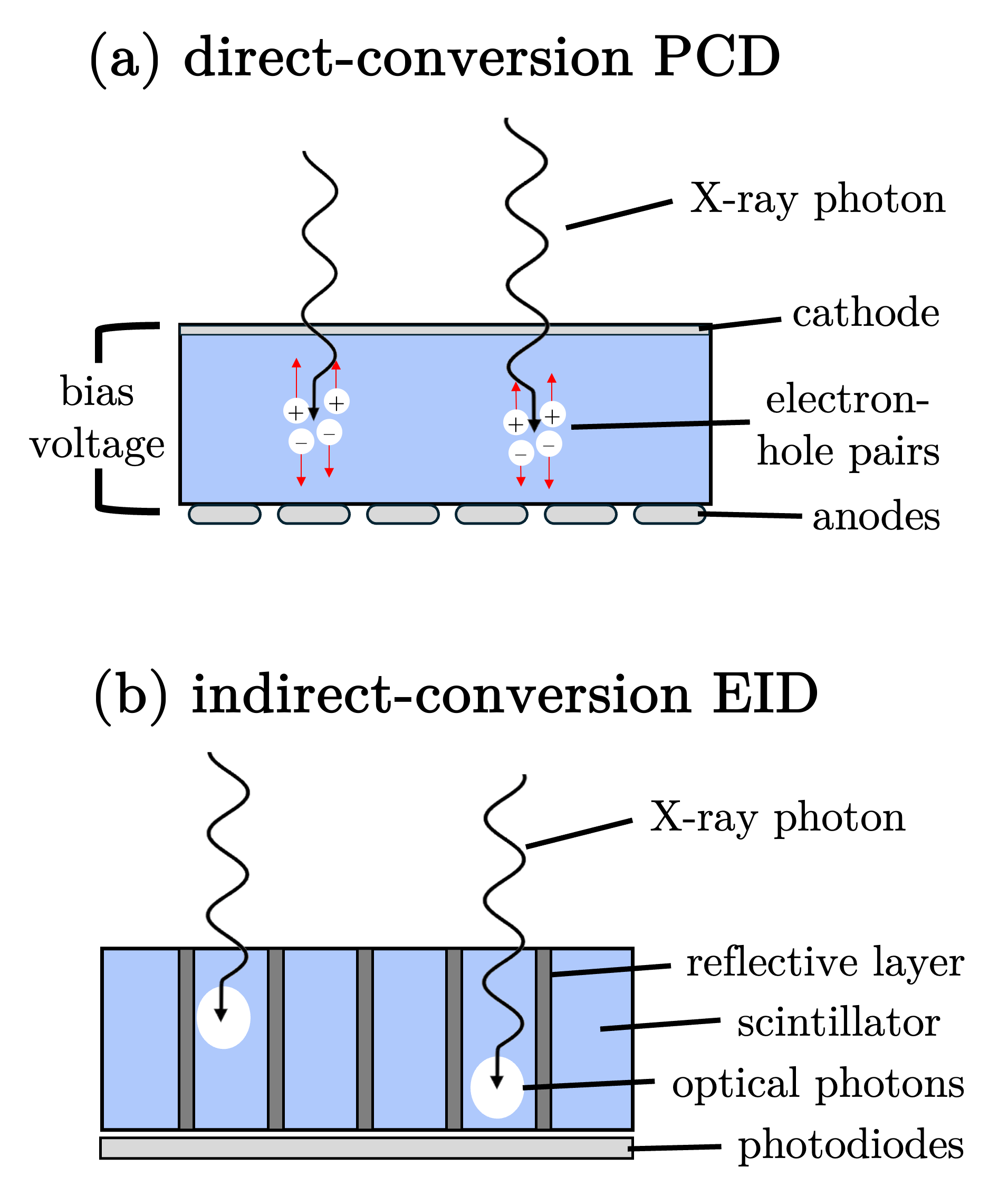
I compared MV-kV dual-energy CT using new photon-counting detectors (PCDs) versus conventional energy-integrating detectors (EIDs), optimizing spectral dose allocation and quantifying contrast-to-noise ratio improvement. Due to the especially severe up-weighting of low-contrast megavoltage X-rays with conventional EIDs, PCDs show particular utility for this application.
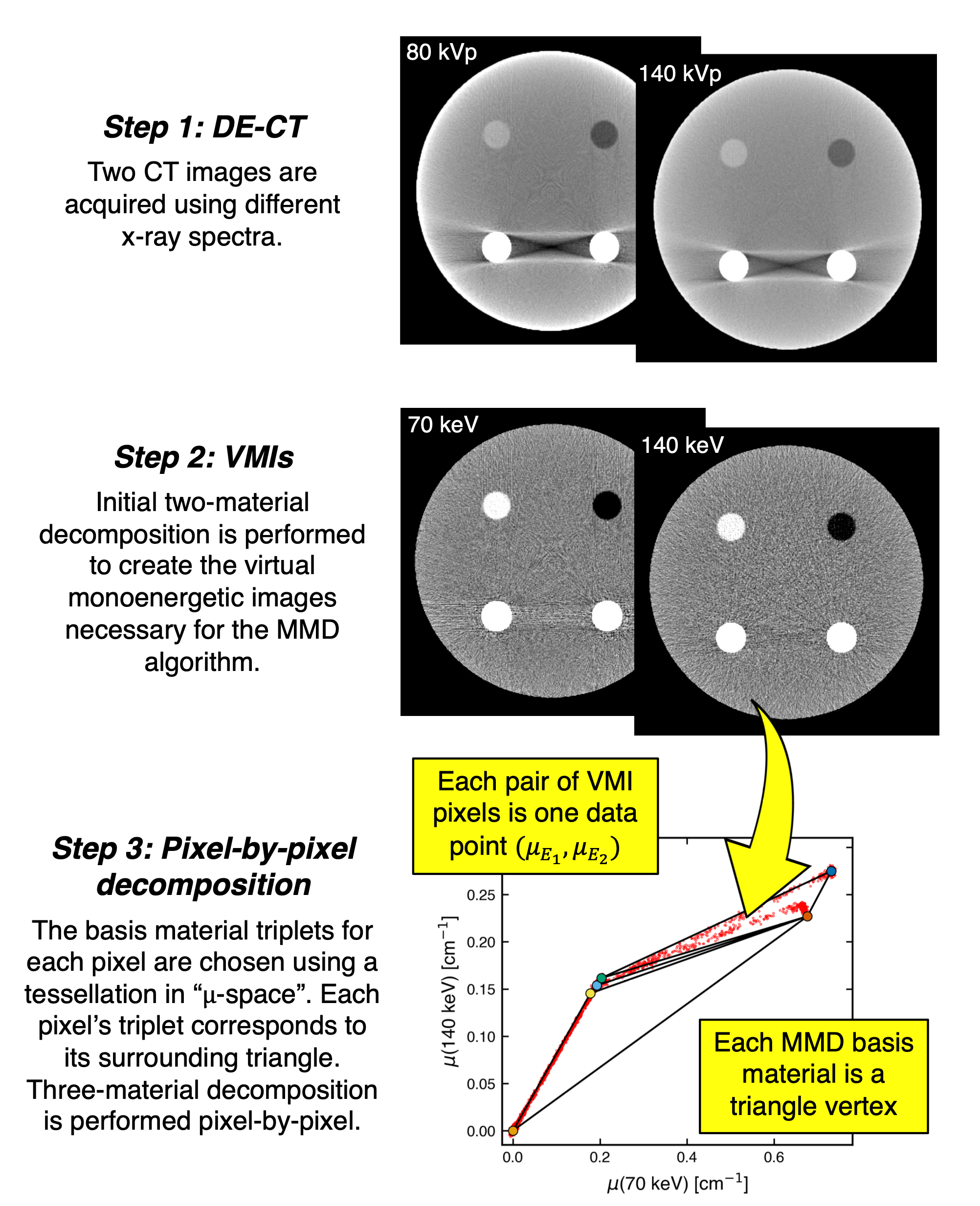
I performed sensitivity analysis of a dual-energy CT method for material decomposition into more than two materials, with applications to plaque risk analysis in cardiac imaging by differentiating soft tissue, adipose, calcium, and iodine. The method utilizes conventional two-material decomposition and a volume constraint to perform material-triplet decomposition, selecting the three basis materials with a pixel-by-pixel criterion.
Get in Touch

giavanna [at] uchicago [dot] edu